Disulfide bonds in protein folding

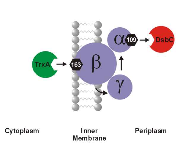
A number of secreted and membrane proteins in both bacteria and eukaryotes contain disulfide bonds. In 1991, we presented evidence that bacteria require the periplasmic protein DsbA for efficient formation of these bonds. DsbA is maintained with its active site cysteines in the oxidized state by the membrane protein DsbB. We are currently carrying out a genetic analysis of DsbA and DsbB to allow a structure-function correlation. The isomerization of incorrectly formed disulfide bonds depends on the periplasmic DsbC protein. This protein, which is maintained with its active site cysteines in the reduced state, receives its electrons from thioredoxin in the cytoplasm via a membrane protein DsbD. We are studying this interesting mechanism of electron transfer across the cytoplasmic membrane.

We are also exploring more fully the role of the various disulfide reducing components of the cytoplasm. These include the thioredoxins and glutaredoxins, but we are also characterizing alternative pathways for disulfide bond reduction.
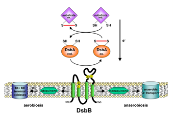
 

Model of the DsbA-DsbB system


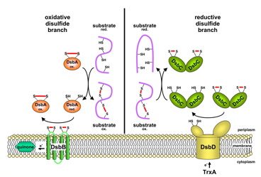
Oxidative and reductive disulfide branch of the E. coli periplasm

Morgan Feeney:

The cytoplasmic redox pathways of E. coli are responsible for the reduction of substrates such as the essential protein ribonucleotide reductase. Two pathways, the glutaredoxin and thioredoxin pathways, function in parallel, with the thioredoxins reduced by thioredoxin reductase (trxB) and the glutaredoxins reduced by glutathione, which is maintained in its reduced state by glutathione reductase (gor). Both pathways ultimately derive their reducing power from NADPH. Although this transfer of electrons down these pathways is known, many questions about the cytoplasmic redox pathways remain unanswered, such as their redundancy and specificity. Suppressor analysis has been used in the past to elucidate these pathways and discover novel biological phenomena. Suppressors of ΔtrxB Δgor are a triplet repeat expansion of ahpC to ahpC*, and in these strains an alkyl hydroxyperoxidase is converted to a disulfide reductase. Because this mutation occurs frequently, we isolated suppressors of ΔtrxB Δgor ΔahpCF in order to discover other pathways for disulfide bond reduction. In these suppressors, we have identified mutations in lpdA, the gene for lipoamide dehydrogenase. LpdA is homologous to Gor and the mutant form of this protein may perform a similar function as Gor in order to reduce disulfide bonds in these suppressors, or may be able to reduce oxidized lipoamide and transfer electrons to ribonucleotide reductase via a novel mechanism.


Hiroshi Kadokura:

Disulfide bonds formed between pairs of cysteines are important structural features of many exported proteins. I have been studying the oxidative branch of disulfide bond formation in the periplasm. My goal is to understand the molecular events that have to occur for the in vivo formation of each disulfide bond in folding proteins.


Rachel Dutton:

Exploring the diversity in redox biology of the bacterial cell envelope

The formation of disulfide bonds is an important event in the folding of many proteins found in the cell envelope of E. coli.  The enzymes that catalyze the formation of disulfide bonds have been identified and characterized in detail in E. coli.  However, there is not a great deal of information regarding this process in other bacteria.  Currently, there are over 500 publicly available genome sequences, representing bacteria from diverse habitats and divergent evolutionary histories.  Using a combination of bioinformatic and experimental approaches, I am interested in addressing the following questions:  Is disulfide bond formation a conserved process across the Domain Bacteria?  If so, are the pathways fully conserved, or are there alternative ways to make disulfide bonds?  If disulfide bond formation is not conserved, which types of bacteria are missing this process? How do these bacteria compensate for a lack of disulfide bonds in protein folding pathways?


Anne-Gaëlle Planson:

Understanding the specificities of E. coli glutaredoxins

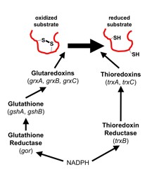
The cytoplasm of Escherichia coli is constantly maintained as a reducing environment in order to allow thousands of specific reactions. The cytoplasmic reduction of disulfide bonds is achieved by the thioredoxin superfamily which includes the glutaredoxin proteins along with glutathione. Members of this protein family are present in most, if not all, living organisms. Glutaredoxins catalyze reversible oxidation/reduction of protein disulfide groups and glutathione-containing mixed disulfide groups. They are involved in different cellular processes such as DNA replication, sulfate assimilation or response to oxidative stress.

The variety of cellular functions performed by the glutaredoxin family is made possible, in part, by the wide range of redox potentials associated with their active site Cys-X-X-Cys motif. Glutaredoxin 1 and glutaredoxin 3 exhibit 33% sequence identity and are structurally nearly superimposable.  Yet, glutaredoxin 1 can reduce the essential enzyme ribonucleotide reductase, which provides deoxyribonucleotides for DNA replication, and PAPS reductase, required for cysteine biosynthesis, while glutaredoxin 3 cannot.  These two proteins provide a good model system for understanding the specificity of these proteins.


Melinda Faulkner

In Escherichia coli, the glutathione/glutaredoxin and thioredoxin pathways are essential for the reduction of cytoplasmic protein disulfide bonds, including those formed in the essential enzyme ribonucleotide reductase during its action on substrates.  Double mutants lacking thioredoxin reductase (trxB) and glutathione reductase (gor) or glutathione biosynthesis (gshA) cannot grow.  Growth of Δgor ΔtrxB strains is restored by a mutant (ahpC*) of the peroxiredoxin AhpC, converting it to a disulfide reductase that generates reduced glutathione.  Our studies show that ahpC* also restores growth to a ΔgshB ΔtrxB strain, which lacks glutathione and accumulates only its precursor γ-glutamylcysteine (γ-Glu-Cys).  Surprisingly, new ahpC suppressor mutations arose in a strain lacking both glutathione and γ-Glu-Cys, ΔgshA ΔtrxB, which ahpC* does not suppress.  Our results show the plasticity of AhpC, which under selective pressure can be altered by different mutations to channel electrons into ribonucelotide reductase by at least four distinct routes, acting via glutathione, γ-Glu-Cys, the thioredoxins or glutaredoxin 1.  The various suppressor strains exhibit an oxidizing cytoplasm and accumulate correctly folded alkaline phosphatase, FAB antibody or tissue plasminogen activator.  Proteins most effectively oxidized vary between strains, potentially providing useful tools for expressing different disulfide-bonded proteins. 

People currently involved in this project:

Hiroshi Kadokura, Markus Eser, Seung Hyun Cho, Melinda Faulkner, Anne-Gaëlle Planson, Rachel Dutton and Morgan Feeney

Recent Publications:

Porat, A., Lillig, C.H., Johansson,C., Fernandes, A.P, Nilsson, L., Holmgren, A., and Beckwith, J. The reducing activity of glutaredoxin 3 towards cytoplasmic substrate proteins is restricted by methionine 43.  Biochemistry 20: 3366-3377 (2007). .

Cho, S.-H., and Beckwith, J. Mutations of the membrane-bound disulfide reductase DsbD that block electron transfer steps from cytoplasm to periplasm in Escherichia coli.  J. Bacteriol. 188:5066-76 (2006).

Gon, S. and Beckwith, J. Ribonucleotide reductases : Influence of environment on synthesis and activity.  Antioxidants and Redox Signalling 8:773-80 (2006).

Gon, S., Faulkner, M.J, and Beckwith, J.  In vivo requirement for glutaredoxins and thioredoxins in the reduction of the ribonucleotide reductases of E. coli. Antioxidants and Redox Signalling 8:735-42(2006).

Segatori, L., Murphy, L., Arredondo, S., Kadokura, H., Gilbert, H., Beckwith, J., and Georgiou, G.  Conserved role of the linker α-helix of the bacterial disulfide isomerase DsbC in the avoidance of misoxidation by DsbB.  J. Biol. Chem.281:4911-9 (2006).

Gon, S., Camara, J., Klungsøyr, H.K., Crooke, E., Skarstad K., and Beckwith, J. Mutations in dnaA and dnaN reveal a novel regulatory mechanism that couples deoxyribonucleotide synthesis and DNA replication during the cell cycle in E. coli. EMBO J. 25:1137-47 (2006).

Sevier, C.S., Kadokura, H., Tam, V.C., Beckwith, J., Fass, D., and Kaiser, C.A. The prokaryotic enzyme DsbB may share structural features with eukaryotic disulfide bond forming oxidoreductases.  Protein Science 14:1630-1642 (2005).

Huber, D., Cha, M.-I., Debarbieux, L., Planson, A.-G., López, G., Tasayco, M.L., Chaffotte, A., Beckwith, J.  A selection for mutants that interfere with folding of E. coli thioredoxin-1 in vivo. Proc. Natl. Acad. Sci., U.S.A 102: 18872-18877 (2005).

Kadokura, H., Nichols, L., and Beckwith, J. Mutational alterations of a key cis proline residue results in the trapping of enzymatic reaction intermediates of DsbA, a member of the thioredoxin superfamily.  J. Bacteriol. 187:1519-1522 (2005).

Berkmen, M., Boyd, D., and Beckwith, J. The non-consecutive disulfide bond of Escherichia coli phytase (AppA) renders it dependent on the protein disulfide isomerase, DsbC. J. Biol. Chem. 280:11387-94 (2005).

Ortenberg R, Gon S, Porat A, Beckwith J. Interactions of glutaredoxins, ribonucleotide reductase, and components of the DNA replication system of Escherichia coli. (2004). Abstract. Paper.

Kadokura H, Tian H, Zander T, Bardwell JC, Beckwith J. Snapshots of DsbA in action: detection of proteins in the process of oxidative folding. (2004). Abstract. Paper.

Katzen F, Beckwith J. Role and location of the unusual redox-active cysteines in the hydrophobic domain of the transmembrane electron transporter DsbD. (2003). Abstract. Paper.

Kadokura H, Katzen F, Beckwith J. Protein Disulfide Bond Formation in Prokaryotes. Annu Rev Biochem. (2003). Abstract. Paper

Haebel PW, Goldstone D, Katzen F, Beckwith J, Metcalf P. The disulfide bond isomerase DsbC is activated by an immunoglobulin-fold thiol oxidoreductase: crystal structure of the DsbC-DsbDalpha complex. EMBO J.21:4774-4784 (2002). Abstract. Paper.

Katzen F, Deshmukh M, Daldal F, Beckwith J. Evolutionary domain fusion expanded the substrate specificity of the transmembrane electron transporter DsbD. EMBO J. 21:3960-3969 (2002). Abstract. Paper.

Kadokura H, Beckwith J. Four cysteines of the membrane protein DsbB act in concert to oxidize its substrate DsbA. EMBO J. 21:2354-63 (2002). Abstract. Paper.

Katzen F, Beckwith J. Disulfide bond formation in the periplasm of Escherichia coli. Methods Enzymol. 348:54-66 (2002). No abstract available.

Ritz D, Beckwith J. Redox state of cytoplasmic thioredoxin. Methods Enzymol. 347:360-70 (2002). No abstract available.

Ritz D, Lim J, Reynolds CM, Poole LB, Beckwith J. Conversion of a peroxiredoxin into a disulfide reductase by a triplet repeat expansion. Science 294:158-160. (2001). Abstract. Paper.

Ritz D, Beckwith J. Roles of thiol-redox pathways in bacteria. Annu. Rev. Microbiol. 55:21-48. (2001). Abstract. Paper.

Kadokura H, Beckwith J. The expanding world of oxidative protein folding. Nat Cell Biol. 3:247-249.(2001). Abstract. Paper.

Goldstone D, Haebel PW, Katzen F, Bader MW, Bardwell JC, Beckwith J, Metcalf, P. DsbC activation by the N-terminal domain of DsbD. Proc. Natl. Acad. Sci. USA. 98:9551-6. (2001). Abstract. Paper.

Katzen F, Beckwith J. Transmembrane Electron Transfer by the Membrane Protein DsbD Occurs via a Disulfide Bond Cascade. Cell. 103:769-779. (2000). Abstract

Kadokura H, Bader M, Tian H, Bardwell JC, Beckwith J. Roles of a conserved arginine residue of DsbB in linking protein disulfide-bond-formation pathway to the respiratory chain of Escherichia coli. Proc. Natl. Acad. Sci. USA. 97:10884-10889. (2000). Abstract. Paper.

Ritz D, Patel H, Doan B, Zheng M, Åslund F, Storz G, Beckwith J. Thioredoxin 2 is involved in the oxidative stress response in Escherichia coli. J. Biol, Chem. 275:2505-2512. (2000). Abstract.

Debarbieux L, Beckwith J. On the functional interchangeability, oxidant vs. reductant, of members of the thioredoxin superfamily. J. Bacteriol. 182:723-727. (2000). Abstract. Paper.

Bessette PH, Åslund F, Beckwith J, Georgiou G. Efficient folding of proteins with multiple disulfide bonds in the Escherichia coli cytoplasm .Proc Natl Acad Sci U S A. 96:13703-13708. (1999). Abstract. Paper.

Stewart E, Katzen F, Beckwith J. Six conserved cysteines of the membrane protein DsbD are required for the transfer of electrons from the cytoplasm to the periplasm of Escherichia coli. EMBO J. 18:5963-5971. (1999). Abstract. Paper.

Debarbieux L, Beckwith J. Electron avenue: Pahtways of disulfide bond formation and isomerization. Cell. 99:117-119. (1999). Review. No abstract available.

Mössner E, Huber-Wunderlich M, Rietsch A, Beckwith J, Glockshuber R, Åslund F. Importance of redox potential for the in vivo function of the cytoplasmic disulfide reductant thioredoxin from Escherichia coli. J. Biol. Chem. 274: 25254-25259(1999) Abstract. Paper.

Åslund F, Zheng M, Beckwith J, Storz G. Regulation of the OxyR transcription factor by hydrogen peroxide and the cellular thiol-disulfide status. Proc Natl Acad Sci U S A. 96:6161-6165(1999) Abstract. Paper.

Åslund F, Beckwith J. Bridge over troubled waters: sensing stress by disulfide bond formation. Cell. 96:751-3 (1999). Review. No abstract available.

Åslund F, Beckwith J. The thioredoxin superfamily: redundancy, specificity, and gray-area genomics. J Bacteriol. 181:1375-9 (1999). Review. Paper.

Rietsch A, Beckwith J. The genetics of disulfide bond metabolism. Annu Rev Genet. 32:163-84 (1998). Review Abstract.

Stewart EJ, Åslund F, Beckwith J. Disulfide bond formation in the Escherichia coli cytoplasm: an in vivo role reversal for the thioredoxins. EMBO J. 17:5543-50 (1998) Abstract. Paper.

Rietsch, A., Bessette, P., Georgiou, G., and Beckwith, J. Reduction of the periplasmic disulfide bond isomerase, DsbC, occurs by passage of electrons from cytoplasmic thioredoxin. J. Bacteriol. 179:6601-6608 (1997). AbstractPaper.

Prinz, W.A., Åslund, F., Holmgren, A., and Beckwith, J. The role of the thioredoxin and glutaredoxin pathways in preventing disulfide bond formation in the cytoplasm. J. Biol. Chem. 272:15661-15667 (1997). AbstractPaper.

Derman, A., Prinz, W., Belin, D., and Beckwith, J. Mutants that allow disulfide bond formation in the cytoplasm of Escherichia coli. Science 262:1744-1746 (1993). Abstract.

Bardwell, J.C.A., McGovern, K., and Beckwith, J. Identification of a protein required for disulfide bond formation in vivo. Cell. 67:581-589 (1991). Abstract.